Val's MRI (Magnetic Resonance Imaging) pictures
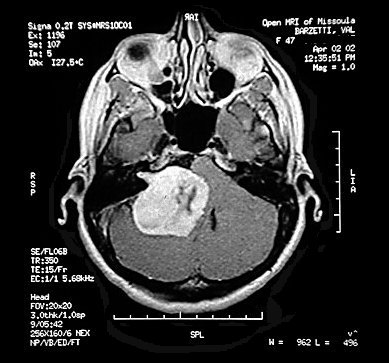
Horizontal MRI section at center of tumor - superior axial view
Note the dark area inside the tumor that may indicate an internal hemorrhage,and the small
extension to the upper left
inside the IAC, the site of origin of the tumor. The tumor has radically deformed
the cerebellum, the part of the brain
that deals with equilibrium and position sense, fine movement, control of muscle tone, and
coordination of muscular activity.
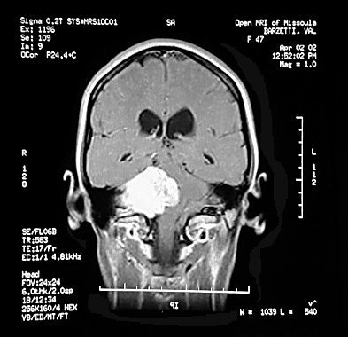
Vertical MRI section at spinal cord - posterior coronal view
Note the severe deflection of the brainstem, a very complex area of the brain.
One part of the brainstem,
the pons, has to do with arousal, wakefulness and alertness (among many other things). In
the pons are
the nuclei for cranial nerves V, VI, VII, and
VIII. Just below the pons is the medulla, which controls breathing,
heartrate, and vasomotor* functions. (*relating to the nerves and muscles that cause the
blood vessels to
constrict or dilate) In the medulla are the nuclei for cranial
nerves VIII, IX, X, XI, and XII.
 5
5
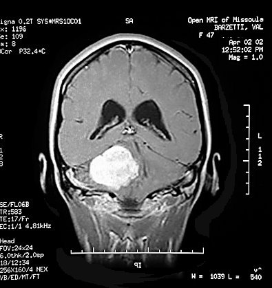
Vertical MRI section 1cm posterior to previous view.
The scales to the side and below the MRI images are in Centimeters. As
measured by the ear surgeon,
Val's tumor is 3.8 x 4 x 5cm. The images have been reverse in the pages to make them
more intuitively
obvious that the tumor is on Val's left side but keep the text readable, so the L/R
indications are now incorrect.