By the ovoid variation (V = 4/3 x Pi x L/2 x W/2 x H/2) for the formula for volume of a sphere (V=4/3 Pi r3) the tumor had a volume of 41.8 cubic centimeters.
Val's 8 month post surgery MRI
images December 4, 2002
| Here are Val's 8 month post-surgery MRI images, put up next to the
"before" images for comparison. I tried to select the ones that were most
similar in cross section location to the first ones. All images have been mirrored so it
is more intuitively obvious that the tumor is on Val's left side, and resized so that the
centimeter scale in the images is the same.
|
|
|
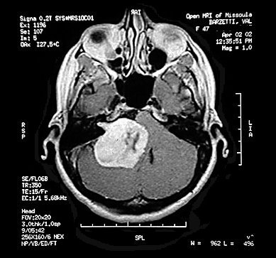
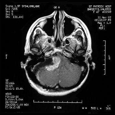
| Before - axial view showing the largest
dimensions of the tumor. The surgeons measured it at 5 x 4.2 x 3.8 centimeters. By the ovoid variation (V = 4/3 x Pi x L/2 x W/2 x H/2) for the formula for volume of a sphere (V=4/3 Pi r3) the tumor had a volume of 41.8 cubic centimeters.
|
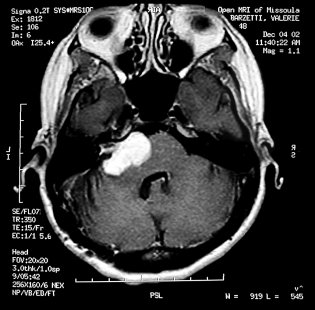
8 months after - axial view also showing the remaining tumor at its largest horizontal dimensions. The surgeon measured it at 3.4 x 2.3 x 2.0 centimeters for a volume of 8.2 cubic centimeters - 19% of it's volume at surgery. |
|
|
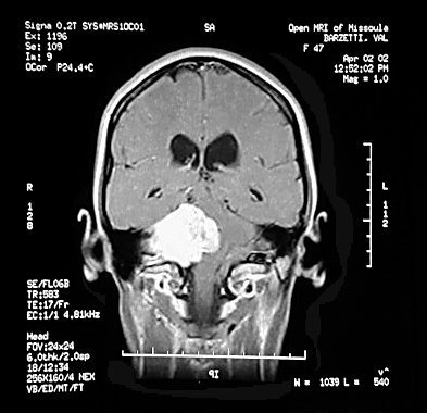
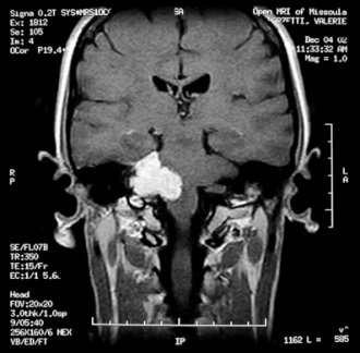
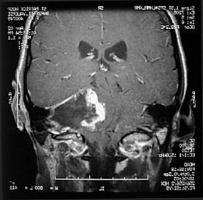
| Before - coronal view Note the severe brainstem deflection. | 8 months after - coronal view The
brainstem has recovered greatly, although somewhat oddly. Val's functioning is fine
however.
|
| Here are the immediate post surgery images for comparison with the 8 month images. | |
|
|
|
|
|